lvh criteria litfl Learn how to diagnose left ventricular hypertrophy (LVH) from ECG voltage and non-voltage criteria, and understand the underlying causes and ECG changes. See examples of LVH with S wave in V2 + R wave in V5 > 35 mm and LV strain pattern. Читайте на сайте Грани.lv свежие новости о происшествиях, интересных событиях и случаях, занимательные курьёзы, интересные факты и конечно же, криминальные новости.
0 · voltage criteria for left ventricular
1 · minimum voltage criteria for lvh
2 · minimal voltage criteria for lvh
3 · lvhn mychart
4 · lvh voltage criteria litfl
5 · left ventricular hypertrophy cornell criteria
6 · criteria for lvh on ekg
7 · calculate lvh
SIA GRIF. VRN 40103003522. Juridiskā adrese: Latgales iela 361, Rīga, LV-1063. Faktiskā adrese: Latgales iela 361, Rīga, LV-1063. Tālr.: 67847637. Fakss: 67187916. E-pasts: [email protected]. Banku konti: SEB banka.
Learn how to diagnose left ventricular hypertrophy (LVH) from ECG voltage and non-voltage criteria, and understand the underlying causes and ECG changes. See examples of LVH with S wave in V2 + R wave in V5 > 35 mm and LV strain pattern.R Wave Peak Time Rwpt - Left Ventricular Hypertrophy (LVH) • LITFL • ECG .ECG Pearl. There are no universally accepted criteria for diagnosing RVH in .
ECG Criteria for Left Atrial Enlargement. LAE produces a broad, bifid P wave in .
For further reading, see LITFL: Sgarbossa Criteria; LBBB with AF: Note the .U Waves - Left Ventricular Hypertrophy (LVH) • LITFL • ECG Library DiagnosisLeft Axis Deviation - Left Ventricular Hypertrophy (LVH) • LITFL • ECG .
Voltage criteria for left ventricular hypertrophy. Deep narrow Q waves < 40 . Learn how to diagnose hypertrophic cardiomyopathy (HCM) based on ECG criteria, such as voltage, Q waves, T waves and WPW. See examples of classic and apical HCM .Learn how to interpret ECG changes in LVH, a common cause of cardiac hypertrophy. Find out the most used indexes, such as Sokolow-Lyon, Cornell and Romhilt-Este, and their sensitivity and specificity.Oct 22, 2024. Home LITFL Eponym. Maurice Sokolow (1911-2002) was an American Cardiologist and educator. Sokolow was known for his contributions to ECG interpretation and .
Learn about the causes, clinical findings, and ECG criteria of left ventricular hypertrophy (LVH), a common cardiac condition. Find out how echocardiography and cardiac .
voltage criteria for left ventricular

Learn the criteria for ECG diagnosis of LVH based on Cornell, Romhilt-Estes, Sokolow-Lyon and other methods. See the points, findings and examples for each criterion. There are several different voltage criteria for diagnosis LVH. Some of the more popular ones are listed below. Remember, voltage criteria alone are not diagnostic of LVH. Voltage criteria for LVH [2]: Limb Leads: R .
dionysus suede shoulder bag dupe
The most commonly used diagnostic criteria for left ventricular hypertrophy (LVH) are based on measurements of QRS voltages.Echocardiographic analysis showed a significant difference in ejection fraction, indexed LVH, and mitral inflow E-wave and A-wave ratio (Table 3). ECG analysis of the test cohort showed that the S waves in leads V 3 and V 4 were good . The Peguero-Lo Presti criteria are novel electrocardiographic (ECG) diagnostic criteria for the detection of left ventricular hypertrophy (LVH) and represent the sum of the .
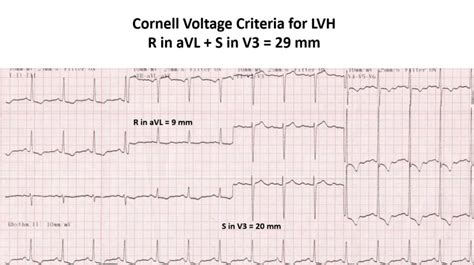
Criteria for ECG Diagnosis of Left Ventricular Hypertrophy. Criterion. Finding. Points. Cornell. Men: V3 S wave + aVL R wave > 28 mm. N/A. Women: V3 S wave + aVL R wave > 20 mm. N/A. Romhilt-Estes (5 points = definite LVH; 4 points = probable LVH) So, looking back at our EKG, it seems like he may meet STEMI criteria. But the voltage on this EKG is awfully high. There are several different voltage criteria for diagnosis LVH. Some of the more popular ones are listed . QRS duration > 100 ms in the presence of a supraventricular rhythm. Most commonly due to bundle branch block or left ventricular hypertrophy. The most important life-threatening causes of QRS widening .
minimum voltage criteria for lvh
LITFL. Cardiac biomarkers (CCC) Buttner R. OMI: Replacing the STEMI misnomer. LITFL 2021; Journal articles. Chew DP, Scott IA, Cullen L, et al. National Heart Foundation of Australia and Cardiac Society of Australia and New Zealand: Australian clinical guidelines for the management of acute coronary syndromes 2016. The Medical journal of . Diagnostic criteria. The QRS is said to be low voltage when: . Co-creator of the LITFL ECG Library. Twitter: @rob_buttner. One comment Aruni Kalawila. May 26, 2020 / 15:14 Reply. Regarding ECG #2, can we diagnose Wellens when there is q waves and loss of R wave progression? Leave a ReplyCancel reply.
ECG Criteria for Left Atrial Enlargement. LAE produces a broad, bifid P wave in lead II (P mitrale) and enlarges the terminal negative portion of the P wave in V1.. In lead II. Bifid P wave with > 40 ms between the two peaks Morphology of ST Depression. ST depression can be either upsloping, downsloping, or horizontal. Horizontal or downsloping ST depression ≥ 0.5 mm at the J-point in ≥ 2 contiguous leads indicates myocardial ischaemia (according to the 2007 Task Force Criteria).Upsloping ST depression in the precordial leads with prominent De Winter T waves is highly specific for .
Voltage criteria for left ventricular hypertrophy (LVH) — S wave in V1 + R wave in V5 > 35mm (note there are many different ways of measuring . Co-creator of the LITFL ECG Library. Twitter: @rob_buttner. 2 Comments Adan R Atriham. December 2, 2021 / 14:02 Reply. Awesome cases, great reviews. I am happy to say that I got all the cases .
This is an example of Pseudo-Wellens syndrome due to left ventricular hypertrophy. ECG Review. LVH by voltage criteria (SV1 + RV6 > 35mm) The pattern of inverted and biphasic T waves is different to Wellens syndrome, affecting multiple leads (i.e. any lead with a tall R wave) rather than V2-3 RWPT in wide QRS complex tachycardia. R-wave peak time (RWPT) may be useful in differentiating ventricular tachycardia (VT) from supraventricular tachycardia (SVT) in patients with wide QRS complex tachycardia:. RWPT duration is measured in lead II from the onset of QRS depolarization until the first change of polarity (with both positive or negative .

Left ventricular hypertrophy (LVH) refers to an increase in the size of myocardial fibers in the main cardiac pumping chamber. Such hypertrophy is usually the response to a chronic pressure or volume load. . the ECG relate to its moderate sensitivity or specificity depending upon which of the many proposed sets of diagnostic criteria are . Smith-Modified Sgarbossa Criteria. As discussed in this article by Stephen Smith, the Smith modified Sgarbossa criteria for Occlusion Myocardial Infarction (OMI) in LBBB have been created to improve diagnostic accuracy. The most important change is the modification of the rule for excessive discordance.. The use of a 5 mm cutoff for excessive discordance was .
American Cardiologist known for his development of ECG criteria for left ventricular hypertrophy (Sokolow-Lyon criteria) Jeremy Rogers and Mike Cadogan . LITFL Top 100 ECG. John Larkin; March 25, 2019; ECG Case 041. 70-year old patient presenting with acute pulmonary oedema. . August 3, 2018; Left Ventricular Hypertrophy (LVH) A review of .
minimal voltage criteria for lvh

LITFL Further Reading. ECG Library Basics – Waves, Intervals, Segments and Clinical Interpretation; ECG A to Z by diagnosis – ECG interpretation in clinical context; ECG Exigency and Cardiovascular Curveball .The sum is 41 mm which is more than 35 mm and therefore LVH is present according to the Sokolow-Lyon criteria. Most commonly used criteria; R in V5 or V6 + S in V1 >35 mm. Cornell Criteria. R in aVL and S in V3 >28 mm in men; . The ventricular complex in left ventricular hypertrophy as obtained by unipolar precordial and limb leads. Am Heart .

ECG Diagnostic criteria. QRS duration > 120ms; RSR’ pattern in V1-3 (“M-shaped” QRS complex) Wide, slurred S wave in lateral leads (I, aVL, V5-6) RBBB: Right Bundle Branch Block . Co-creator of the LITFL ECG .However, the criteria are very specific (i.e., specificity >90%, which means if the criteria are met, it is very likely that ventricular hypertrophy is present). Left Ventricular Hypertrophy (LVH) General ECG features include: ≥ QRS amplitude (voltage criteria; i.e., tall R-waves in LV leads, deep S-waves in RV leads)
Left ventricular hypertrophy (LVH) is thickening of the heart muscle of the left ventricle of the heart, . The Cornell criteria for LVH are: S in V 3 + R in aVL > 28 mm (men) S in V 3 + R in aVL > 20 mm (women) The Romhilt-Estes point score system ("diagnostic" >5 .
ECG criteria. Left axis deviation (usually -45 to -90 degrees) . In LAFB, the QRS voltage in lead aVL may meet voltage criteria for LVH (R wave height > 11 mm), but there will be no LV strain pattern. . LITFL Further Reading. ECG Library Basics – Waves, Intervals, . LVH is recognized as causing many false-positive cardiac catheterization lab activations. The electrocardiographic diagnostic criteria for LVH are numerous, but poorly sensitive. The most widely used criteria is the Sokolow-Lyon criteria: If the sum of the amplitude of the S in V1 + R in V5/6 >35 mm, LVH is present. Patterns of Myocardial Ischaemia Two main ECG patterns associated with NSTEACS: ST segment depression; T wave flattening or inversion; While there are numerous conditions that may simulate myocardial ischaemia (e.g. left ventricular hypertrophy, digoxin effect), dynamic ST segment and T wave changes (i.e. different from baseline ECG or .
gucci chain bag dupe
References. Zema MJ, Kligfield P. ECG poor R-wave progression: review and synthesis. Arch Intern Med. 1982 Jun;142(6):1145-8. [PMID 6212033] References. Sovari AA, Farokhi F, Kocheril AG. Inverted U wave, a specific electrocardiographic sign of cardiac ischemia. Am J Emerg Med. 2007 Feb;25(2):235-7.LITFL ECG library is a free educational resource covering over 100 ECG topics relevant to Emergency Medicine and Critical Care. All our ECGs are free to reproduce for educational purposes, provided: The image is credited to litfl.com. The teaching activity is on a not-for-profit basis. The image is not otherwise labelled as belonging to a third . The most commonly used diagnostic criteria for left ventricular hypertrophy (LVH) are based on measurements of QRS voltages. The ECG criteria for LVH shown in Table 1 have evolved over the years. 65–78 Criteria were originally based on R and S amplitudes in standard limb leads I and III, using clinical and autopsy data as reference standards. 4–6 .
chloe shoulder bag dupe
Wellens Syndrome. Wellens syndrome is a pattern of inverted or biphasic T waves in V2-3 (in patients presenting with/following ischaemic sounding chest pain) that is highly specific for critical stenosis of the left anterior descending artery.. There are two patterns of T-wave abnormality in Wellens syndrome:. Type A = Biphasic T waves with the initial deflection .
EMS-Grivory Grilamid® LV-50H FWA black 9225 Nylon 12, 50% Glass Fiber Filled, Conditioned. Manufacturer Notes: EMS-Grivory (EMS-CHEMIE) Category Notes. Plastic. PA, Polyamide. Some of the values displayed above may have been converted from their original units and/or rounded in order to display the information in a consistent format.
lvh criteria litfl|calculate lvh


