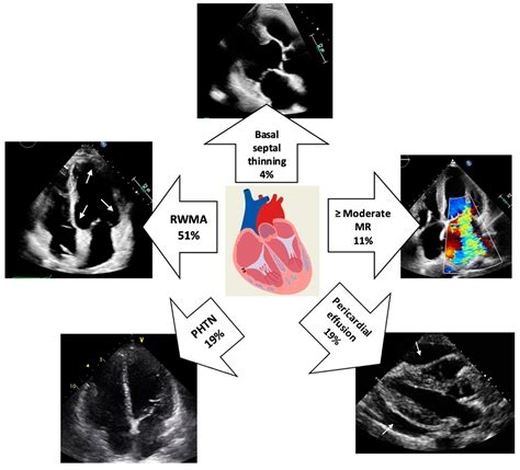
lv rwma means Regional wall motion abnormalities are defined as regional abnormalities in contractile function. Ischemic heart disease is the most common cause of wall . LV Edge 25mm Reversible Belt. With their sleek straps and gleaming signature buckles, Louis Vuitton’s belts for women are chic, versatile – and an indispensable fashion accessory. Made from the Maison’s iconic Monogram or Damier canvases, or from a variety of luxurious leathers, these waist-defining pieces are available in a wide range of .
0 · rwma of heart meaning
1 · rwma echocardiogram left ventricle
2 · rwma echocardiogram
3 · no rwma of the heart
4 · no resting rwma means
Get the best deals for fake louis vuitton at eBay.com. We have a great online selection at the lowest prices with Fast & Free shipping on many items!
rwma of heart meaning
Regional wall motion abnormalities are defined as regional abnormalities in contractile function. Ischemic heart disease is the most common cause of wall . Regional wall motion abnormality on an echocardiogram means that a region of the heart muscle is not contracting as it normally should. Short form often used is RWMA. RWMA is a very common medical abbreviation .
No RWMA means that all segments of the left ventricle are contracting normally. At rest means that no stress test like exercise test or stress test with medications (e.g. dobutamine, adenosine) has been done to check .
dior overshirt
Regional wall motion abnormality means that the motion of a region of the heart muscle is abnormal. It is a term commonly used in echocardiography. Echocardiography is ultrasound imaging of the heart. Very . Regional wall motion abnormalities (WMAs) after myocardial infarction are associated with adverse remodeling and increased mortality in the short to medium term. Their long‐term prognostic impact is less well .Left ventricular RWMA is described as a hypokinesis, dyskinesis, or akinesis of a segment when compared to the other contracting segments of the chamber. This can be visualized sonographically as a blunting of the typical symmetric .Despite the similar improvement in endocardial border delineation, LVO settings allow the detection of more WMA than MCE at peak stress, leading to a significantly higher accuracy for .
Left ventricular wall motion abnormalities are regularly assessed visually on echocardiography and cardiac MRI. The evaluation is primarily based on systolic wall .Assessment of left ventricular systolic function has a central role in the evaluation of cardiac disease. Accurate assessment is essential to guide management and prognosis. Numerous echocardiographic techniques are used in the .
Three-dimensional echocardiography provides a volumetric measurement of global and regional left ventricular (LV) function. It avoids the subjectivity of 2D echocardiography in the . What does it mean by “no resting RWMA of heart”? Is a question which comes to the mind of many who read an echocardiogram report for the first time.RWMA is s.
: https://johnsonfrancis.org/general/what-does-it-mean-by-no-resting-rwma-of-heart/ RWMA is short for regional wall motion abnormality, a terminology used.The aim of this study is to compare between conventional 2DE and 3DE in the detection of RWMA and evaluation of LV regional and global function. 2. . regional EF of the hypokinetic areas was comparable to that of the akinetic areas. This means that RT3DE could not differentiate between hypokinesia and akinesia. Table 5. Comparison between 2DE . The aim of this study is to compare between conventional 2DE and 3DE in the detection of RWMA and evaluation of LV regional and global function. 2 . regional EF of the hypokinetic areas was comparable to that of the akinetic areas. This means that RT3DE could not differentiate between hypokinesia and akinesia. Table 5. Comparison between 2DE .To evaluate the usefulness of echocardiographic regional wall motion abnormalities (RWMA) in detecting coronary artery disease (CAD) in patients with left ventricular (LV) dysfunction and a normal-sized or dilated left ventricle, 103 patients were studied by two-dimensional echocardiography (2DE) and cardiac catheterization.
where t is the time from negative dp/dt, P 0 the mono-exponential amplitude coefficient, P τ the mono-exponential time constant, and P ∞ is the mono-exponential asymptotic equation.. AoDP and AoSP were also measured after pulling out the catheter from the left ventricle to the ascending aorta. LVEDP, AoDP, and AoDP were recorded as average values of . The TTC cohort showed higher LV diastolic and systolic volumes, a lower LV EF, and a higher WMSI than ant-STEMI patients (Table 2). RWMA involving the apex with sparing of the base were detected in 29% and 2% of patients with TTC and ant-STEMI, respectively (P = 0.002). Right ventricular involvement and LV outflow tract obstruction were more .
Regional wall motion abnormality means that the motion of a region of the heart muscle is abnormal. It is a term commonly used in echocardiography. Echocardiography is ultrasound imaging of the heart. Very often echocardiography reports use the short form RWMA instead of the full form regional wall motion abnormality.
Assessment of left ventricular systolic function has a central role in the evaluation of cardiac disease. Accurate assessment is essential to guide management and prognosis. Numerous echocardiographic techniques are used in the assessment, each with its own advantages and disadvantages. This review is based on a literature search of the PubMed, MEDLINE, .
Cardiac wall motion abnormalities describe kinetic alterations in the cardiac wall motion during the cardiac cycle and have an effect on cardiac function.Cardiac wall motion abnormalities can be categorized with respect to their degree and their distribution pattern that is whether they are global or segmental and whether they can be attributed to a coronary . Introduction. Left ventricular (LV) dysfunction is a serious condition in the critically ill patient. This can cause low cardiac output and cardiovascular instability, leading to hypoperfusion of vital organs and contributing to multi . Background Regional Wall Motion Abnormality (RWMA) serves as an early indicator of myocardial infarction (MI), the global leader in mortality. Accurate and early detection of RWMA is vital for the successful treatment of MI. Current automated echocardiography analyses typically concentrate on peak values from left ventricular (LV) displacement curves, .raphy; and presented RWMA in two or more adjacent segments of the LV. Exclusion criteria were age <18 years, a history of heart fail-ure, CAD, or cardiomyopathy. Echocardiographic reports were manually reviewed to identify patients with RWMA. The LV regional function was evaluated using the 17-segment heart model, and segments were marked as .

In the intensive care unit (ICU), point-of-care ultrasound (POCUS) has emerged as an invaluable tool for clinicians. POCUS enhances bedside clinical decision-making, ensures the safety of critical procedures, and monitors therapeutic intervention outcomes. 1 The roles of the left ventricle (LV) and right ventricle (RV) are pivotal in the physiology of shock states, a . The main sign of ischemia during stress echocardiography (SE) is the transient regional wall motion abnormality (RWMA) [] due to flow-limiting coronary artery disease (CAD).RWMA can be provoked by exercise or pharmacological stressors [2, 3].All main SE modalities (exercise, dobutamine, vasodilators) have comparable accuracy for the diagnosis of . RWMA – Echo. The term regional wall motion abnormality or RWMA can be used in any imaging which shows movements of the myocardial segments like echocardiography, cine cardiac magnetic resonance imaging, cine computed tomography and nuclear cardiology imaging. The left ventricular myocardium has been divided into 17 segments by the American .
Within the test cohort, the DL model accurately identified any RWMA with an area under the curve of 0.96 (0.92-0.98). The mean F1 scores of the experts and the DL model were numerically similar for 6 of 7 regions: anterior (86 vs 84), anterolateral (80 vs 74), inferolateral (83 vs 87), inferoseptal (86 vs 86), apical (88 vs 87), inferior (79 vs 81), and any RWMA (90 vs . Collectively hypovolemia, RV/LV systolic failure, LV RWMA, and valve abnormalities represent the main culprits for hemodynamic and respiratory deterioration. 15, 19, 21 As some patients may have more than one cause of deterioration, by identifying unrecognized findings, rTEE compensates for potentially missed causes when failure of initial .
Impairment of left ventricular (LV) diastolic function is common amongst those with left heart disease and is associated with significant morbidity. Given that, in simple terms, the ventricle can only eject the volume with which it fills and that approximately one half of hospitalisations for heart failure (HF) are in those with normal/’preserved’ left ventricular .

very irresistible givenchy sephora
The stitching on a Louis Vuitton belt is a crucial element to examine when determining its authenticity. Genuine belts feature even, precise stitching that is almost invisible. Counterfeit belts often have sloppy stitching with uneven thread and loose ends.
lv rwma means|no resting rwma means


