lvh strain Left ventricular hypertrophy (LVH): Markedly increased LV voltages: huge precordial R and S waves that overlap with the adjacent leads (SV2 + RV6 >> 35 mm). R-wave peak time > 50 ms in V5-6 with associated QRS broadening. LV strain pattern with ST . This chic shoulder bag is crafted of classic Louis Vuitton monogram on toile canvas, with textured black leather trim. The bag features a polished brass chain-link wristlet strap and an optional toffee brown leather shoulder strap. The top zipper opens to a black fabric interior with a patch pocket. 1319303.
0 · normal lv strain echo values
1 · minimum voltage criteria for lvh
2 · minimal voltage for lvh
3 · lvh with strain pattern meaning
4 · lvh with strain pattern
5 · lvh with strain ekg
6 · lv strain normal values
7 · how to assess lv function
The Coffee Bean & Tea Leaf, Las Vegas: See unbiased reviews of The Coffee Bean & Tea Leaf, one of 5,522 Las Vegas restaurants listed on Tripadvisor.
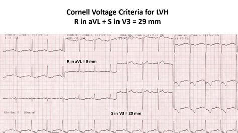
Left ventricular hypertrophy (LVH): Markedly increased LV voltages: huge precordial R and S waves that overlap with the adjacent leads (SV2 + RV6 >> 35 mm). R-wave peak time > 50 ms in V5-6 with associated QRS broadening. LV strain pattern with ST .
RWPT in wide QRS complex tachycardia. R-wave peak time (RWPT) may be .Right ventricular strain pattern with T-wave inversion and ST depression in the right .
ECG Criteria for Left Atrial Enlargement. LAE produces a broad, bifid P wave in .References. Da Costa D, Brady WJ, Edhouse J. Bradycardias and .
References. Sovari AA, Farokhi F, Kocheril AG. Inverted U wave, a specific .Left Axis Deviation = QRS axis less than -30°.. Normal Axis = QRS axis between . LVH with strain pattern can sometimes be seen in long standing severe aortic regurgitation, usually with associated left ventricular hypertrophy and systolic dysfunction. The sensitivity of LVH strain pattern on ECG as a .The most common causes of left ventricular hypertrophy are aortic stenosis, aortic regurgitation, hypertension, cardiomyopathy and coarctation of the aorta. There are several ECG indexes, which generally have high diagnostic .
Left ventricular hypertrophy (LVH) refers to an increase in the size of myocardial fibers in the main cardiac pumping chamber. Such hypertrophy is usually the response to a .
Left ventricular hypertrophy, or LVH, is a term for a heart’s left pumping chamber that has thickened and may not be pumping efficiently. Sometimes problems such as aortic stenosis or high blood pressure overwork . To diagnose left ventricular hypertrophy, a healthcare professional does a physical exam and asks questions about your symptoms and family's health history. The care .Left ventricular hypertrophy (LVH) makes it harder for the heart to pump blood efficiently. It can result in a lack of oxygen to the heart muscle. It can also cause changes to the heart’s .
Electrocardiographic (ECG) left ventricular hypertrophy (LVH) with strain pattern is said to be present when, apart from the voltage criterion for ECG-LVH, there is also a downsloping .
This is referred to as “LVH with strain” or “LVH with repolarization abnormality.” At times, these repolarization abnormalities can mimic ischemic ST changes, and distinguishing them from. Left ventricular hypertrophy (LVH) refers to an increase in the size of myocardial fibers in the main cardiac pumping chamber. Such hypertrophy is usually the response to a chronic pressure or volume load. Ogah OS, Oladapo OO, Adebiyi AA, et al. Electrocardiographic left ventricular hypertrophy with strain pattern: prevalence, mechanisms and prognostic implications. Cardiovascular Journal of Africa . January-February . To diagnose left ventricular hypertrophy, a healthcare professional does a physical exam and asks questions about your symptoms and family's health history. The care professional checks your blood pressure and listens to your heart with a device called a stethoscope. . They can improve blood flow and decrease the strain on the heart. Side .
Left ventricular hypertrophy (LVH) doesn’t usually cause symptoms at first, but when symptoms do occur, they often mirror those in conditions such as heart failure. A physical exam and cardiac . BackgroundBoth ECG strain pattern and QRS measured left ventricular (LV) hypertrophy criteria are associated with LV hypertrophy and have been used for risk stratification. However, the independent predictive value of ECG strain in apparently healthy individuals in predicting mortality and adverse cardiovascular events is unclear. Methods and ResultsMESA . The classic left ventricular (LV) strain pattern of ST segment depression and T-wave inversion on the left precordial leads of the standard resting ECG is a well-known marker of the presence of anatomic LV hypertrophy (LVH). 1–6 Furthermore, the occurrence of this electrocardiographic abnormality of ventricular repolarization has been associated with a .
orologio cartier crash
V4-V6 precordial leads may show ST depression & T wave inversions known as the LV Strain pattern; Romhilt-Estes Criteria. Diagnostic ≥ 5 points and probable ≥ 4 points) ECG Criteria: . ↑ Sokolow M, Lyon TP: The ventricular complex in left ventricular hypertrophy as obtained by unipolar precordial and limb leads. Am Heart J 37: 161, 1949 It appears that our patient meets LVH voltage criteria. With LVH, it is common to see a strain pattern, where there are ST Depression and T Wave inversions in the left sided leads [2]. Is this patient having LVH with strain or is it a true STEMI? Armstrong et al introduced a flow chart to help identify a STEMI in patients with LVH [5]: Electrocardiographic left ventricular hypertrophy (LVH) has many faces with countless features. Beyond the classic measures of LVH, including QRS voltage and duration, the left ventricular (LV) strain pattern is an element whereby characteristic R-ST depression is followed by a concave ST segment that ends in an asymmetrically inverted T wave.Electrocardiographic left ventricular hypertrophy with strain pattern has been documented as a marker for left ventricular hypertrophy. Its presence on the ECG of hypertensive patients is associated with a poor prognosis. This review was undertaken .
Left ventricular hypertrophy (LVH) is a condition in which there is an increase in left ventricular mass, either due to an increase in wall thickness or due to left ventricular cavity enlargement, or both. Most commonly, the left ventricular wall thickening occurs in response to pressure overload, and chamber dilatation occurs in response to the volume overload.[1] Left Ventricular Hypertrophy With Strain. Submitted by Dawn on Thu, 12/12/2013 - 10:38. This ECG is from a man with left ventricular hypertrophy. LVH causes taller-than-normal QRS complexes in leads oriented toward the left side of the heart, such as Leads I, II, aVL, V4, V5, and V6. Leads on the opposite side, such as V1, V2, and V3, will have . Left Ventricular Hypertrophy With Strain. Submitted by Dawn on Thu, 12/12/2013 - 10:38. This ECG is from a man with left ventricular hypertrophy. LVH causes taller-than-normal QRS complexes in leads oriented toward the left side of the heart, such as Leads I, II, aVL, V4, V5, and V6. Leads on the opposite side, such as V1, V2, and V3, will have .

Left ventricular hypertrophy (LVH) is a well‐known risk factor for cardiovascular morbidity and mortality. 1, 2, 3 Early diagnosis is essential as regression of LVH is associated with a reduction in cardiovascular events. 4, 5, 6 Although several modalities are available for detection of LVH, ECG and echocardiography are most commonly used in . It is now appreciated that electrocardiographic LVH with ST-segment and T-wave abnormalities occurs in conditions that are not necessarily caused by increased hemodynamic work, as in patients with dilated or hypertrophic cardiomyopathies, and that lesser degrees of ST-T abnormalities than the “typical strain” pattern are associated with LVH . Electrical remodeling of the left ventricle in the setting of hypertrophy causes ST elevation in leads V1-V3, as well as the "strain" pattern. In patients with LVH, this elevation in leads V1-V3 may mimic an anterior .
Left Ventricular Hypertrophy (LVH) causes tall R waves in left sided and deep S waves in right sided leads. Always look for ‘strain’ pattern in left sided leads. . 3 points – for ST segment and T wave changes (‘typical . Electrocardiographic left ventricular hypertrophy (LVH) has many faces with countless features. Beyond the classic measures of LVH, including QRS voltage and duration, the left ventricular (LV) strain pattern is an element whereby characteristic R-ST depression is followed by a concave ST segment that ends in an asymmetrically inverted T wave. Esimerkiksi vasemman kammion lihasseinämän paksuntumisen eli hypertrofian (LVH) ns. Sokolow-Lyon-kriteeri (kytkennän V1 alaspäin suuntautuvan eli S-heikahduksen ja V5 ylöspäin suuntautuvan eli R-heilahduksen summa) on yli 35 mm, mutta tämän tarkkuutta lisää huomattavasti liitännäismuutosten (ST-T eli ”strain” -muutos) huomioon . The classic strain pattern of ST depression and T-wave inversion on the ECG is a well recognized marker of the presence of anatomic left ventricular hypertrophy (LVH), 1–7 independent of the possible relationship of this repolarization abnormality to underlying coronary heart disease. 7 Indeed, the presence of even minimal degrees of ST depression in the lateral .
Myocardial strain is a dimensionless variable representing the change in length between two points over the cardiac cycle, and can be quantified using echocardiography or CMR tissue tracking. Regional strain is less reproducible; whereas peak global strain is more consistent, supporting the use of global longitudinal strain (GLS) in clinical .Left ventricular hypertrophy (LVH) is a major risk factor for cardiovascular disease, including sudden death. Antihypertensive therapy in patients with LVH results in lowering of blood pressure and drug-dependent reduction of LV mass. Complete reversal of LVH can be.Left ventricular (LV) hypertrophy (LVH) is a frequent imaging finding in daily clinical practice, and its presence is associated with poor outcomes and ventricular arrhythmias. It is commonly detected in athletes, arterial hypertension, aortic stenosis, .
In electrocardiography, a strain pattern is a well-recognized marker for the presence of anatomic left ventricular hypertrophy (LVH) in the form of ST depression and T wave inversion on a resting ECG. [1] It is an abnormality of repolarization and it has been associated with an adverse prognosis in a variety heart disease patients. It has been important in refining the role of ECG . Introduction. The strain pattern in the 12‐lead ECG, defined as ST‐segment depression and T‐wave inversion, represents ventricular repolarization abnormalities.1 The mechanism underlying ECG strain is unclear, although it has been proposed as subendocardial ischemia.2, 3 ECG strain is associated with concentric left ventricular (LV) hypertrophy (LVH), . Prevalences of echocardiographic LVH, abnormal LV geometry and LV midwall function in patients with or without ECG strain are shown in Table 3. In this population selected on the basis of ECG LVH by Cornell product and/or Sokolow-Lyon voltage, strain was associated with a higher prevalence of LVH defined by LV mass/body surface area in patients with and .
Assessment of left ventricular systolic function has a central role in the evaluation of cardiac disease. Accurate assessment is essential to guide management and prognosis. Numerous echocardiographic techniques are used in the assessment, each with its own advantages and disadvantages. This review is based on a literature search of the PubMed, MEDLINE, .
normal lv strain echo values

About Press Copyright Contact us Creators Advertise Developers Terms Privacy Policy & Safety How YouTube works Press Copyright Contact us Creators Advertise .
lvh strain|how to assess lv function


